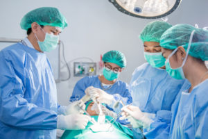
General Surgery

Surgery is a medical specialty that uses operative techniques to investigate or treat a pathological condition. Besides the focus on surgical techniques, pre-operative assessment, post-operative care and management of both surgical and medical complications are also important.


Traditionally surgery meant making a large incision to perform a procedure which may result in more harm. With the advance in technology, minimally invasive surgery becomes more popular. Common surgical procedures included tumour resection, skin and subcutaneous mass excision, laparoscopic and endoscopic surgery, hernia repair surgery, cholecystectomy, varicose vein surgery and thyroidectomy etc.

Surgery

Skin and subcutaneous mass


Skin and subcutaneous masses such as sebaceous cyst, lipoma, naeves and warts are very common. The mass is excised during operation and sent for pathological examination. Many of these operations could be performed under local anaesthesia.

Colectomy


Large bowel is the last part of digestive system. Water is absorbed in large bowel and remaining waste material is stored as faeces before being removed by defecation. The large bowel is divided into caecum, ascending colon, transverse colon, descending colon, sigmoid colon, rectum and anal canal.


Colectomy is performed for cases of colorectal cancer, large colonic polyp, inflammation including diverticulitis or inflammatory bowel disease or bowel infarction. CT scan of abdomen is performed before operation to identify the location of the disease or stage of the colorectal cancer. It could be done either by open surgery (large abdominal incision) or laparoscopic surgery, depends on the tumour size and your health condition. The diseased part colon is resected, and anastomosis or stoma is performed resection. Laparoscopic surgery will result in smaller incision, faster recovery and less postoperative complications when compared with open surgery.

Hernioplasty (Hernia repair surgery)


Hernia occurs when an organ (usually small intestine, large intestine) or intra-abdominal fat protrudes trough a weak point in abdominal wall, which result in a swelling in abdominal wall. If you have increased pressure within abdomen, chronic cough, strenuous activity, you may have a higher chance to have hernia. Common sites of hernia included inguinal region, umbilicus or post-operative wounds. Hernia commonly presents as a swelling over abdominal wall which disappears when you lie down. Surgery is advised as there is a chance of strangulation of hernia content which is a life-threatening condition. When there is severe pain and persistent swelling even when you lie down, emergent surgery is indicated.

Gastrectomy


Gastrectomy is a partial or total surgical resection of stomach. Types of gastrectomy included total gastrectomy, partial gastrectomy or sleeve gastrectomy etc. It is recommended if you have stomach cancer, gastric or duodenal ulcers with severe bleeding or perforation, or morbid obesity.

Hepatectomy


Liver located at right upper quadrant of your abdomen. It consists of two lobes, both right and left lobe. Its main job is to filter the blood coming from the digestive tract, before passing to the rest of your body. The liver also detoxifies chemicals and metabolizes drugs. Hepatectomy is surgical resection of the liver. It is performed for both benign and malignant hepatic tumour. It is also performed for tumour in gallbladder or bile duct, intrahepatic gallstones or parasitic cysts of the liver.

Esophagectomy


Esophagus is a muscular tube which connects the throat to the stomach. It travels behind the trachea and heart, passes through the diaphragm and empties into the stomach. Esophagectomy is a surgical resection of part or whole esophagus with reconstruction afterwards. It is performed for esophageal cancer, Barrett’s esophagus (pre-cancerous condition), esophageal perforation or esophageal stricture.

Cholecystectomy


Gallbladder is a pear-shaped organ at right upper abdomen, just under your liver. It stores bile which helps break down fat in food. Gallbladder diseases such as gallstones and inflammation of gallbladder are common. Cholecystectomy (removal of gallbladder) is suggested if you have frequent pain due to the gallstone or in case of gallbladder inflammation. It is also performed if you have gallbladder polyp, inflammation in bile duct or pancreas due to gallstone disease or cancer of gallbladder. This surgery can be done by laparoscopic method which results in smaller wounds, less post-operative pain and faster recovery.

Anal fistula


Anal fistula is a chronic abnormal communication between the anal canal and the perianal skin. It commonly occurs in patient with history of anal abscess which does not heal properly. It will cause perianal pain and swelling or pus discharge over perianal region.

Appendicectomy


This is a surgery performed for appendicitis. Appendicitis is an inflammation of the appendix. Appendix is a blind end tube at the junction of small intestine and large intestine which is found at right lower abdomen. You will have pain at right lower abdominal pain, vomiting and fever. Emergent surgery is needed once the diagnosis is made. If left untreated, appendix may rupture and peritonitis will occur which is fatal.

Haemorrhoidectomy


Haemorrhoid are vascular structures in anal canal. They act as cushions which help stool control normally. They become a disease when get swollen or inflamed. They will cause bright red rectal bleeding during or after defacation, mucus discharge, perianal mass that prolapse through anus, itchiness or perianal pain. If you have chronic constipation, low fiber diet, prolonged sitting, increased intra-abdominal pressure such as pregnancy, you may have higher chance to develop hemorrhoidal disease.


Haemorrhoids could be divided into internal haemorrhoids and external haemorrhoids

Internal haemorrhoids could be classified into different grade according to their severity

Grade I: No prolapse, just prominent blood vessels

Grade II: Prolapse upon bearing down, but spontaneous reduction

Grade III: Prolapse upon bearing down requiring manual reduction

Grade IV: Prolapse with inability to be manually reduced


Management for grade I and grade II haemorrhoid:

  • High fiber diet
  • Topical cream or suppositories
  • Sclerotherapy
  • Rubber band ligation


Rubber band ligation:

This can be performed in clinic. Doctor will introduce an instrument (proctoscope) into your anus and examine the severity of the internal haemorrhoid. Rubber band ligation will be performed if there is grade II internal haemorrhoids. Suction is applied onto the haemorrhoid and a rubber band is applied onto it. The band will cause necrosis of the haemorrhoid. After procedure, you may have perianal discomfort or minimal perianal pain which may lasted for few hours to few days. You may also have small amount of per-rectal bleeding, especially after 10-14 days after banding when the banding sloughs off.


Management for grade III and grade IV haemorrhoid:

  • Open haemorrhoidectomy
  • Stapler haemorrhoidopexy


Open haemorrhoidectomy:

  • Performed for both internal and external haemorrhoid
  • Diseased haemorrhoids are excised, which result in wounds over perianal region
  • Operative time: 30-45 mins
  • There will be wound pain after operation and may last for 1-2 weeks. The post – operative pain is well controlled by analgesic
  • Perianal wounds will heal around 6-8 weeks after operation


Stapler haemorrhoidopexy:

  • Abnormally enlarged haemorrhoidal tissues are excised, followed by repositioning of the remaining hemorrhoidal tissue back to its normal anatomical position
  • Operative time: 30-45 mins
  • Not suitable for external haemorrhoids or case of anal stenosis
  • Recommended for grade 3 or 4 internal haemorrhoids
  • Less post-operative pain and there is no wound over perianal region

Thyroidectomy


Thyroid gland is at the anterior neck, just below the Adam’s apple. The thyroid gland secretes hormones which control your body’s metabolic rate. It consists of two lobes connected by an isthmus. Hemithyroidectomy is the resection of one thyroid lobe. Total thyroidectomy is the removal of whole thyroid gland. These operations are performed for cases including thyroid nodules, thyroid cancer and goitre. This surgery could be performed by traditional open surgery (incision made over anterior neck) or endoscopic method.

Circumcision


Foreskin is the fold of skin that covers the head of penis. By the age of five years old, most boys can pull back their foreskin. If the foreskin is too long or too tight, it will cover the urethral opening and it may cause difficulty during urination and problem in cleaning of penis. In these circumstances, circumcision is indicated. Circumcision is a surgical removal of a sleeve of foreskin and the wound is closed with sutures or stapler. The surgery can be performed in clinic or hospital under local or general anesthesia.

Ingrown toenail


Ingrown toenail occurs when the edges or corners of the nails grow into the skin next to the nail. It commonly occurs at the first toe. If you always wear ill-fitting shoes, have excessive sweating or poor nail care, your nail will curl and dig into the skin, resulting in ingrown toenail which cause pain, swelling or even inflammation. Surgery is required in case of severe pain and inflammation.


Causes of ingrown toenail:

  • Poor nail care, included cutting the nail too short, rounded off at the tip or peeled off at the edges instead of being cut straight across.
  • Wear ill-fitting shoes that are too narrow or too short
  • Poor foot hygiene or excessive sweating
  • Trauma to nail plate or toe
  • Bacterial infection over nail or toe

Surgery for varicose veins


Varicose veins are dilated and tortuous veins which commonly found in lower limbs. These abnormally dilated veins are caused by malfunction of the valves in the veins. Healthy valves maintain a single direction flow of blood from feet back to the heart. However, there may be backflow of blood in the veins if these valves malfunction. These may cause aching or cramp like pain over lower limbs. If left untreated, they may cause pigmentation, bleeding or ulcer over lower limbs.


Traditional Surgery – ligation and stripping

This operation is done under general anesthesia. An incision is made at the groin area and the varicose vein is ligated at the junction where it meets the deep vein. After the ligation, the varicose vein is stripped out. Smaller incisions are made over the calf for avulsion of smaller varicose veins.


Minimal invasive surgery

This surgery results in smaller wounds, less post-operative pain and better cosmetic results.

  • Radiofrequency ablation (RFA)
    The varicose vein is identified and marked with ultrasound. A small incision is made at thigh region over the varicose vein and a laser fiber probe is inserted into the vein. Then radiofrequency energy is released which heat up the wall of the varicose vein. The energy is applied continuously along the whole varicose vein, causing the ablation of the vein.


  • Sclerosant or foaming agent injection
    Sclerosant or foaming agent is injected into the varicose vein under ultrasound guidance. This can seal the vein and the vein will disappear in few weeks. This procedure is suitable smaller size dilated veins such as spider veins and reticular veins.

Breast Surgery

Breast lesion biopsy


There are two types of breast biopsy, fine needle aspiration and core biopsy. Smaller needle is used for fine needle aspiration. For core biopsy, larger sized needle is used, and more tissues can be sampled. Both procedures can be performed under local anesthesia. The breast lesion is localized either by palpation or ultrasound, then local anesthesia is injected at the entry site of the needle. The needle will be inserted into the lesion through the skin and tissue is sampled for examination. Usually there will be minimal pain and bruises over the biopsy site.

Breast mass excision (lumpectomy)


Breast mass could be either benign (eg. Fibroadenoma) or malignant. If the mass is big, symptomatic or suspicious of malignancy, excision is suggested. This operation is performed under general anesthesia. Incision is made at circumareolar region and the mass is excised. The mass will be sent for histological examination afterwards.

Breast conservative surgery


This is one of the surgical treatments for breast cancer. The breast cancer is excised with adequate margins and axillary lymph node sampling (sentinel lymph node dissection) is also performed. Sentinel lymph nodes is the first group of lymph nodes to which cancer cells are most likely to spread from the breast cancer. Radioactive substance or dye will be injected around the breast tumour and sentinel lymph nodes will be localized and excised. If there is cancer cell in the sentinel lymph node, axillary dissection will be done. You must receive radiotherapy to the breast tumour resection site after operation in order to provide better local disease control and lower local recurrence rate. You can preserve your breast after breast conservative surgery, however, there may be a possibility of second operation if there is cancer cell found at the resection margin.

Mastectomy


This is another surgical treatment for breast cancer. The whole breast including the cancer is excised. After surgery, there is transverse scar over chest wall. Nipple is resected during same operation. This may cause psychological burden for some patients.

Breast reconstruction


After removal of the breast cancer, creation of new breast with tissue from another part of your body (back or lower abdomen) or implant can be done. This could be performed immediately after breast surgery or months or years later when you finish all the breast cancer treatments. This surgery will have better cosmetic outcomes and less psychological burdens for breast cancer patients.

Endoscopy and endoscopic treatments

A flexible fiber optic scope is introduced into your body to examine the interior of hallow organ. Commonly performed endoscopies included esophagogastroduodenoscopy, colonoscopy and sigmoidoscopy. These endoscopies are usually performed under anesthesia. Scope will be introduced through mouth or anus and the gastrointestinal tract will be distended with air and thorough examination could be done. Biopsy, removal of polyps, control bleeding over ulcers could also be performed with the endoscopy. After the procedure, you could be discharged and resumed your normal activities.


Esophagogastroduodenoscopy (OGD)


The endoscopy is introduced into your throat through your mouth and the scope will be advanced to esophagus, stomach down to second part of duodenum. If there is ulcers, polyps or tumor, biopsy or polypectomy (removal of polyp) can be performed. Tissue from stomach will be taken for helicobacter infection also. Helicobacter infection is commonly seen infection in upper gastrointestinal tract, and this will cause epigastric
discomfort, epigastric pain and peptic ulcers and this will also increase the risk for gastric cancer if left untreated. If you have epigastric pain, acid reflux/heartburn, swallowing difficulty, your doctor will arrange OGD for further investigation.

A flexible fiber optic scope is introduced into your body to examine the interior of hallow organ. Commonly performed endoscopies included esophagogastroduodenoscopy, colonoscopy and sigmoidoscopy. These endoscopies are usually performed under anesthesia. Scope will be introduced through mouth or anus and the gastrointestinal tract will be distended with air and thorough examination could be done. Biopsy, removal of polyps, control bleeding over ulcers could also be performed with the endoscopy. After the procedure, you could be discharged and resumed your normal activities.


Colonoscopy


This can directly visualize the large intestine and can perform polypectomy and biopsy in same session. The scope is introduced through your anus and air is inflated into the bowel. After distension of the bowel, scope is advanced from anal canal up to caecum (the first part of large intestine). Banding or sclerotherapy for haemorrhoid can be done at the end of the examination.

A flexible fiber optic scope is introduced into your body to examine the interior of hallow organ. Commonly performed endoscopies included esophagogastroduodenoscopy, colonoscopy and sigmoidoscopy. These endoscopies are usually performed under anesthesia. Scope will be introduced through mouth or anus and the gastrointestinal tract will be distended with air and thorough examination could be done. Biopsy, removal of polyps, control bleeding over ulcers could also be performed with the endoscopy. After the procedure, you could be discharged and resumed your normal activities.


Capsule endoscopy


It contains a tiny camera and an array of LEDS powered by a battery. After you swallow the capsule, it passes along your gastrointestinal tract and takes number of images per second. The images are transmitted wirelessly to a receiver which connected to a portable recording device. It passes through esophagus, stomach, duodenum, small bowel, large bowel and pass out your body through anus during defecation. After the completion of the examination, nurse will collect your portable recording device and doctor will review the images. This is especially useful for examination of small bowel pathology (such as polyps, ulcers, abnormal vascular structures or tumour) which may cause abdominal pain, unexplained gastrointestinal bleeding or anaemia.

A flexible fiber optic scope is introduced into your body to examine the interior of hallow organ. Commonly performed endoscopies included esophagogastroduodenoscopy, colonoscopy and sigmoidoscopy. These endoscopies are usually performed under anesthesia. Scope will be introduced through mouth or anus and the gastrointestinal tract will be distended with air and thorough examination could be done. Biopsy, removal of polyps, control bleeding over ulcers could also be performed with the endoscopy. After the procedure, you could be discharged and resumed your normal activities.


Endoscopic retrograde cholangiopancreatography (ERCP)


This combines the use of endoscopy and fluoroscopy to diagnose and treat biliary or pancreatic ductal diseases. If you have biliary tract obstruction by stones, structures, tumour, your doctor will perform ERCP for removal of stones or insertion of stent to relieve the obstruction. The endoscopy enters your body through your mouth and is advanced down to second part of duodenum. Contrast is injected into the biliary tree through the bile duct opening at second part of duodenum. Biliary tract obstruction will be seen under Xrays. Biliary tract stone can be removed, and stent can be passed into biliary tract if there is any obstruction. Tissue biopsy could also be obtained during the procedure.

EJ Medical Specialist Centre
Suite 1412, 14/F,

Ocean Centre, Harbour City,

5 Canton Road, TST, HK


Tel : +852 2396 3038